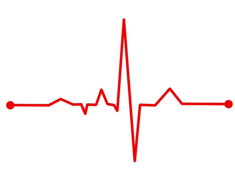
What is junctional rhythm ecg? This is a question that many people have heard of, but don’t know the answer to. Junctional rhythm is an ECG reading that occurs when the heart’s electrical activity happens in the junction between the atria and ventricles. In this article, we will discuss what junctional rhythm is, what causes it, and how it can be treated.
The heart is responsible for pumping blood throughout the body. In order to do this, it uses a series of electrical signals that cause the heart to contract in certain parts of its beating cycle. These signals begin in an area called the sinoatrial node, or S-A node, which is located near the top of your right atrium. These electrical signals travel from the S-A node to another part of your heart called the atrioventricular node, or A-V node. The A-V node is located between your right atrium and right ventricle. From the A-V node, these electrical signals then travel into a group of cells known as the ventricles. This is where the heart pumps blood throughout the body.
Although it is responsible for moving blood, your heart is mainly composed of muscle tissue and circulatory systems that are similar to those found in other organs in your body. But unlike other parts of your body, your heart’s valves work very differently; they have three flaps to a single orifice and function as independently functioning units.
Your heart has two major chambers: the right atrium, which receives deoxygenated blood from your body through veins, and the left atrium, which receives oxygenated blood from your lungs through your pulmonary artery. Each chamber is divided into smaller sections by valves that let blood flow in only one direction. The right and left atria are separated by the interatrial septum, a thin membrane; the two chambers are separated by the interventricular septum, another thin barrier.
The heart also has four valves that help it pump blood efficiently to your body: the tricuspid valve on the right side allows blood to flow out of the right atrium into the right ventricle; the pulmonary valve lets blood flow from your right ventricle into your lungs’ pulmonary artery; and the mitral and aortic valves between your left atrium and left ventricle let blood out of these chambers.
Your heart also has a rhythm of its own. Your heart’s pacemaker, called the sinus node or sinoatrial node, is located just beneath the right atrium in your upper chest. The pacemaker generates electrical impulses that travel out from this node throughout your heart, causing it to contract and pump blood efficiently through your body. And if your heart ever needs a little extra help, your blood vessels have it. Your blood vessels regulate the amount of blood reaching your heart during each heartbeat based on how much oxygen it needs.
We hope this information on junctional rhythm ecg was helpful.


